

Which Change Would Lead To A Decrease In The Expression Of A Gene?

Regulation of cistron expression by a hormone receptor
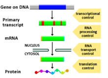
Diagram showing at which stages in the Deoxyribonucleic acid-mRNA-protein pathway expression can be controlled
Regulation of gene expression, or gene regulation,[1] includes a broad range of mechanisms that are used past cells to increment or decrease the product of specific cistron products (protein or RNA). Sophisticated programs of gene expression are widely observed in biology, for example to trigger developmental pathways, respond to environmental stimuli, or adapt to new food sources. Virtually any stride of gene expression can be modulated, from transcriptional initiation, to RNA processing, and to the post-translational modification of a protein. Oftentimes, one cistron regulator controls some other, and so on, in a gene regulatory network.
Gene regulation is essential for viruses, prokaryotes and eukaryotes equally it increases the versatility and adjustability of an organism by allowing the jail cell to limited poly peptide when needed. Although equally early on every bit 1951, Barbara McClintock showed interaction betwixt ii genetic loci, Activator (Air conditioning) and Dissociator (Ds), in the color germination of maize seeds, the first discovery of a gene regulation system is widely considered to exist the identification in 1961 of the lac operon, discovered by François Jacob and Jacques Monod, in which some enzymes involved in lactose metabolism are expressed past E. coli only in the presence of lactose and absence of glucose.
In multicellular organisms, cistron regulation drives cellular differentiation and morphogenesis in the embryo, leading to the creation of dissimilar jail cell types that possess unlike gene expression profiles from the same genome sequence. Although this does not explain how gene regulation originated, evolutionary biologists include it as a fractional caption of how evolution works at a molecular level, and information technology is central to the science of evolutionary developmental biology ("evo-devo").
Regulated stages of gene expression [edit]
Any stride of cistron expression may be modulated, from signaling to transcription to post-translational modification of a protein. The post-obit is a listing of stages where gene expression is regulated, the near extensively utilized point is Transcription Initiation:
- Betoken transduction
- Chromatin, chromatin remodeling, chromatin domains
- Transcription
- Post-transcriptional modification
- RNA transport
- Translation
- mRNA degradation
Modification of Dna [edit]
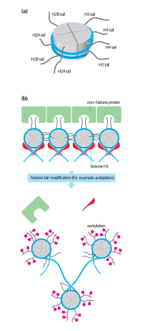
Histone tails and their function in chromatin formation
In eukaryotes, the accessibility of big regions of DNA can depend on its chromatin structure, which can be contradistinct as a upshot of histone modifications directed by Deoxyribonucleic acid methylation, ncRNA, or Deoxyribonucleic acid-bounden protein. Hence these modifications may upwards or downwardly regulate the expression of a gene. Some of these modifications that regulate cistron expression are inheritable and are referred to as epigenetic regulation.
Structural [edit]
Transcription of DNA is dictated by its structure. In full general, the density of its packing is indicative of the frequency of transcription. Octameric poly peptide complexes chosen histones together with a segment of DNA wound effectually the eight histone proteins (together referred to as a nucleosome) are responsible for the amount of supercoiling of Dna, and these complexes can be temporarily modified past processes such every bit phosphorylation or more permanently modified by processes such as methylation. Such modifications are considered to exist responsible for more than or less permanent changes in gene expression levels.[two]
Chemical [edit]
Methylation of DNA is a mutual method of gene silencing. DNA is typically methylated by methyltransferase enzymes on cytosine nucleotides in a CpG dinucleotide sequence (also chosen "CpG islands" when densely amassed). Assay of the blueprint of methylation in a given region of DNA (which can be a promoter) tin can be achieved through a method called bisulfite mapping. Methylated cytosine residues are unchanged by the treatment, whereas unmethylated ones are changed to uracil. The differences are analyzed past Dna sequencing or past methods adult to quantify SNPs, such as Pyrosequencing (Biotage) or MassArray (Sequenom), measuring the relative amounts of C/T at the CG dinucleotide. Aberrant methylation patterns are thought to exist involved in oncogenesis.[3]
Histone acetylation is also an important process in transcription. Histone acetyltransferase enzymes (HATs) such as CREB-binding protein also dissociate the Dna from the histone complex, assuasive transcription to proceed. Often, Deoxyribonucleic acid methylation and histone deacetylation work together in gene silencing. The combination of the 2 seems to be a signal for Deoxyribonucleic acid to exist packed more densely, lowering gene expression.[ citation needed ]
Regulation of transcription [edit]
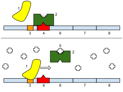
1: RNA Polymerase, 2: Repressor, iii: Promoter, iv: Operator, 5: Lactose, half dozen: lacZ, 7: lacY, 8: lacA. Top: The factor is essentially turned off. There is no lactose to inhibit the repressor, so the repressor binds to the operator, which obstructs the RNA polymerase from bounden to the promoter and making lactase. Bottom: The gene is turned on. Lactose is inhibiting the repressor, allowing the RNA polymerase to bind with the promoter, and limited the genes, which synthesize lactase. Eventually, the lactase will digest all of the lactose, until at that place is none to bind to the repressor. The repressor will then bind to the operator, stopping the industry of lactase.
Regulation of transcription thus controls when transcription occurs and how much RNA is created. Transcription of a gene by RNA polymerase tin can exist regulated past several mechanisms. Specificity factors alter the specificity of RNA polymerase for a given promoter or set of promoters, making it more or less likely to bind to them (i.east., sigma factors used in prokaryotic transcription). Repressors bind to the Operator, coding sequences on the DNA strand that are close to or overlapping the promoter region, impeding RNA polymerase'south progress forth the strand, thus impeding the expression of the factor. The image to the correct demonstrates regulation by a repressor in the lac operon. General transcription factors position RNA polymerase at the start of a protein-coding sequence so release the polymerase to transcribe the mRNA. Activators enhance the interaction between RNA polymerase and a particular promoter, encouraging the expression of the gene. Activators do this by increasing the attraction of RNA polymerase for the promoter, through interactions with subunits of the RNA polymerase or indirectly by changing the construction of the Dna. Enhancers are sites on the DNA helix that are bound by activators in social club to loop the DNA bringing a specific promoter to the initiation circuitous. Enhancers are much more common in eukaryotes than prokaryotes, where only a few examples exist (to date).[four] Silencers are regions of DNA sequences that, when bound by particular transcription factors, can silence expression of the gene.
Regulation past RNA [edit]
RNA can be an of import regulator of cistron activity, e.chiliad. by microRNA (miRNA), antisense-RNA, or long non-coding RNA (lncRNA). LncRNAs differ from mRNAs in the sense that they have specified subcellular locations and functions. They were first discovered to be located in the nucleus and chromatin, and the localizations and functions are highly diverse now. Some still reside in chromatin where they interact with proteins. While this lncRNA ultimately affects gene expression in neuronal disorders such every bit Parkinson, Huntington, and Alzheimer disease, others, such as, PNCTR(pyrimidine-rich non-coding transcriptors), play a role in lung cancer. Given their role in illness, lncRNAs are potential biomarkers and may be useful targets for drugs or gene therapy, although there are no approved drugs that targert lncRNAs nonetheless. There number of lncRNAs in the homo genome remains poorly defined, but some estimates range from 16,000 to 100,000 lnc genes.[5]
Epigenetic Gene Regulation [edit]

Overview of Epigenetic mechanisms.
Epigenetics refers to the modification of genes that is not changing the DNA or RNA sequence. Epigenetic modifications are also a key gene in influencing gene expression. They occur on genomic Dna and histones and their chemical modifications regulate cistron expression in a more than efficient manner. There are several modifications of DNA (usually methylation) and more than 100 modifications of RNA in mammalian cells." Those modifications event in altered protein binding to DNA and a modify in RNA stability and translation efficiency.[6]
Special cases in human biology and illness [edit]
Regulation of transcription in cancer [edit]
In vertebrates, the majority of gene promoters comprise a CpG island with numerous CpG sites.[7] When many of a cistron'southward promoter CpG sites are methylated the gene becomes silenced.[eight] Colorectal cancers typically have three to 6 driver mutations and 33 to 66 hitchhiker or passenger mutations.[9] Still, transcriptional silencing may exist of more than importance than mutation in causing progression to cancer. For example, in colorectal cancers virtually 600 to 800 genes are transcriptionally silenced by CpG island methylation (see regulation of transcription in cancer). Transcriptional repression in cancer can as well occur by other epigenetic mechanisms, such as altered expression of microRNAs.[10] In breast cancer, transcriptional repression of BRCA1 may occur more than frequently by over-expressed microRNA-182 than by hypermethylation of the BRCA1 promoter (run across Low expression of BRCA1 in breast and ovarian cancers).
Regulation of transcription in addiction [edit]
One of the central features of addiction is its persistence. The persistent behavioral changes announced to be due to long-lasting changes, resulting from epigenetic alterations affecting gene expression, inside particular regions of the encephalon.[11] Drugs of corruption cause three types of epigenetic alteration in the encephalon. These are (one) histone acetylations and histone methylations, (2) Dna methylation at CpG sites, and (3) epigenetic downregulation or upregulation of microRNAs.[11] [12] (Encounter Epigenetics of cocaine addiction for some details.)
Chronic nicotine intake in mice alters brain cell epigenetic command of gene expression through acetylation of histones. This increases expression in the brain of the protein FosB, important in addiction.[13] Cigarette addiction was besides studied in about 16,000 humans, including never smokers, electric current smokers, and those who had quit smoking for upwardly to 30 years.[xiv] In blood cells, more than eighteen,000 CpG sites (of the roughly 450,000 analyzed CpG sites in the genome) had frequently altered methylation amidst current smokers. These CpG sites occurred in over 7,000 genes, or roughly a third of known human genes. The bulk of the differentially methylated CpG sites returned to the level of never-smokers within five years of smoking cessation. Nevertheless, 2,568 CpGs among 942 genes remained differentially methylated in erstwhile versus never smokers. Such remaining epigenetic changes can be viewed as "molecular scars"[12] that may touch cistron expression.
In rodent models, drugs of corruption, including cocaine,[15] methamphetamine,[16] [17] booze[18] and tobacco smoke products,[19] all crusade DNA damage in the brain. During repair of Deoxyribonucleic acid amercement some individual repair events can alter the methylation of Dna and/or the acetylations or methylations of histones at the sites of harm, and thus can contribute to leaving an epigenetic scar on chromatin.[xx]
Such epigenetic scars likely contribute to the persistent epigenetic changes constitute in addiction.
Regulation of transcription in learning and memory [edit]

DNA methylation is the addition of a methyl group to the Deoxyribonucleic acid that happens at cytosine. The image shows a cytosine unmarried ring base and a methyl group added on to the 5 carbon. In mammals, DNA methylation occurs most exclusively at a cytosine that is followed by a guanine.
In mammals, methylation of cytosine (see Effigy) in DNA is a major regulatory mediator. Methylated cytosines primarily occur in dinucleotide sequences where cytosine is followed by a guanine, a CpG site. The total number of CpG sites in the man genome is approximately 28 million.[21] and generally almost lxx% of all CpG sites have a methylated cytosine.[22]
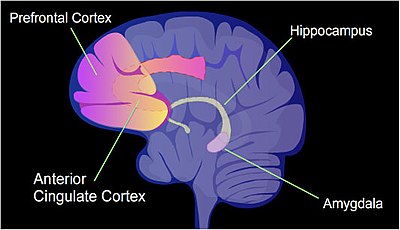
The identified areas of the human brain are involved in memory formation.
In a rat, a painful learning experience, contextual fear workout, can result in a life-long fearful memory later a single training event.[23] Cytosine methylation is altered in the promoter regions of nearly 9.17% of all genes in the hippocampus neuron Deoxyribonucleic acid of a rat that has been subjected to a brief fear conditioning experience.[24] The hippocampus is where new memories are initially stored.
Methylation of CpGs in a promoter region of a gene represses transcription[25] while methylation of CpGs in the body of a gene increases expression.[26] TET enzymes play a central role in demethylation of methylated cytosines. Demethylation of CpGs in a gene promoter by TET enzyme activeness increases transcription of the gene.[27]
When contextual fear workout is applied to a rat, more than 5,000 differentially methylated regions (DMRs) (of 500 nucleotides each) occur in the rat hippocampus neural genome both ane 60 minutes and 24 hours after the conditioning in the hippocampus.[24] This causes near 500 genes to be upwards-regulated (often due to demethylation of CpG sites in a promoter region) and about 1,000 genes to be downwards-regulated (oft due to newly formed v-methylcytosine at CpG sites in a promoter region). The design of induced and repressed genes inside neurons appears to provide a molecular footing for forming the first transient retention of this training event in the hippocampus of the rat brain.[24]
Post-transcriptional regulation [edit]
After the DNA is transcribed and mRNA is formed, there must exist some sort of regulation on how much the mRNA is translated into proteins. Cells exercise this past modulating the capping, splicing, addition of a Poly(A) Tail, the sequence-specific nuclear export rates, and, in several contexts, sequestration of the RNA transcript. These processes occur in eukaryotes but not in prokaryotes. This modulation is a outcome of a protein or transcript that, in turn, is regulated and may have an analogousness for certain sequences.
Three prime untranslated regions and microRNAs [edit]
Three prime number untranslated regions (3'-UTRs) of messenger RNAs (mRNAs) often contain regulatory sequences that mail-transcriptionally influence gene expression.[28] Such 3'-UTRs often contain both binding sites for microRNAs (miRNAs) as well every bit for regulatory proteins. By binding to specific sites within the iii'-UTR, miRNAs can decrease gene expression of various mRNAs by either inhibiting translation or directly causing deposition of the transcript. The 3'-UTR also may have silencer regions that bind repressor proteins that inhibit the expression of a mRNA.
The 3'-UTR oftentimes contains miRNA response elements (MREs). MREs are sequences to which miRNAs bind. These are prevalent motifs within iii'-UTRs. Among all regulatory motifs inside the 3'-UTRs (due east.m. including silencer regions), MREs make upwards nearly half of the motifs.
Equally of 2014, the miRBase web site,[29] an annal of miRNA sequences and annotations, listed 28,645 entries in 233 biologic species. Of these, 1,881 miRNAs were in annotated human miRNA loci. miRNAs were predicted to have an average of about four hundred target mRNAs (affecting expression of several hundred genes).[thirty] Freidman et al.[thirty] judge that >45,000 miRNA target sites within human being mRNA 3'-UTRs are conserved above groundwork levels, and >60% of human protein-coding genes have been under selective force per unit area to maintain pairing to miRNAs.
Direct experiments bear witness that a unmarried miRNA tin reduce the stability of hundreds of unique mRNAs.[31] Other experiments show that a single miRNA may repress the production of hundreds of proteins, simply that this repression often is relatively mild (less than two-fold).[32] [33]
The effects of miRNA dysregulation of factor expression seem to be important in cancer.[34] For example, in gastrointestinal cancers, a 2015 newspaper identified nine miRNAs as epigenetically altered and constructive in downwards-regulating Deoxyribonucleic acid repair enzymes.[35]
The furnishings of miRNA dysregulation of gene expression also seem to be of import in neuropsychiatric disorders, such every bit schizophrenia, bipolar disorder, major depressive disorder, Parkinson's illness, Alzheimer'southward disease and autism spectrum disorders.[36] [37] [38]
Regulation of translation [edit]
The translation of mRNA tin also exist controlled by a number of mechanisms, more often than not at the level of initiation. Recruitment of the small ribosomal subunit can indeed be modulated by mRNA secondary structure, antisense RNA binding, or protein bounden. In both prokaryotes and eukaryotes, a large number of RNA bounden proteins exist, which often are directed to their target sequence by the secondary structure of the transcript, which may alter depending on certain conditions, such as temperature or presence of a ligand (aptamer). Some transcripts act every bit ribozymes and self-regulate their expression.
Examples of gene regulation [edit]
- Enzyme consecration is a process in which a molecule (e.g., a drug) induces (i.east., initiates or enhances) the expression of an enzyme.
- The induction of heat shock proteins in the fruit fly Drosophila melanogaster.
- The Lac operon is an interesting example of how gene expression can be regulated.
- Viruses, despite having only a few genes, possess mechanisms to regulate their gene expression, typically into an early and late phase, using collinear systems regulated by anti-terminators (lambda phage) or splicing modulators (HIV).
- Gal4 is a transcriptional activator that controls the expression of GAL1, GAL7, and GAL10 (all of which lawmaking for the metabolic of galactose in yeast). The GAL4/UAS system has been used in a variety of organisms beyond diverse phyla to report cistron expression.[39]
Developmental biology [edit]
A large number of studied regulatory systems come up from developmental biology. Examples include:
- The colinearity of the Hox cistron cluster with their nested antero-posterior patterning
- Blueprint generation of the paw (digits - interdigits): the gradient of sonic hedgehog (secreted inducing factor) from the zone of polarizing activity in the limb, which creates a slope of active Gli3, which activates Gremlin, which inhibits BMPs likewise secreted in the limb, results in the formation of an alternate pattern of activity every bit a outcome of this reaction–diffusion system.
- Somitogenesis is the creation of segments (somites) from a uniform tissue (Pre-somitic Mesoderm). They are formed sequentially from anterior to posterior. This is accomplished in amniotes possibly by means of two opposing gradients, Retinoic acrid in the anterior (wavefront) and Wnt and Fgf in the posterior, coupled to an oscillating design (segmentation clock) composed of FGF + Notch and Wnt in antiphase.[40]
- Sex activity conclusion in the soma of a Drosophila requires the sensing of the ratio of autosomal genes to sex chromosome-encoded genes, which results in the production of sexless splicing factor in females, resulting in the female person isoform of doublesex.[41]
Circuitry [edit]
Up-regulation and down-regulation [edit]
Up-regulation is a process that occurs within a cell triggered by a signal (originating internal or external to the jail cell), which results in increased expression of one or more genes and as a issue the proteins encoded past those genes. Conversely, downward-regulation is a procedure resulting in decreased cistron and corresponding poly peptide expression.
- Up-regulation occurs, for instance, when a cell is deficient in some kind of receptor. In this instance, more receptor protein is synthesized and transported to the membrane of the cell and, thus, the sensitivity of the cell is brought dorsum to normal, reestablishing homeostasis.
- Down-regulation occurs, for example, when a jail cell is overstimulated past a neurotransmitter, hormone, or drug for a prolonged period of time, and the expression of the receptor protein is decreased in society to protect the cell (run across besides tachyphylaxis).
Inducible vs. repressible systems [edit]

Gene regulation works using operators and repressors in bacteria.
Gene Regulation tin can be summarized by the response of the respective system:
- Inducible systems - An inducible arrangement is off unless in that location is the presence of some molecule (called an inducer) that allows for cistron expression. The molecule is said to "induce expression". The mode by which this happens is dependent on the control mechanisms as well every bit differences between prokaryotic and eukaryotic cells.
- Repressible systems - A repressible system is on except in the presence of some molecule (chosen a corepressor) that suppresses cistron expression. The molecule is said to "repress expression". The manner by which this happens is dependent on the control mechanisms besides as differences between prokaryotic and eukaryotic cells.
The GAL4/UAS system is an example of both an inducible and repressible system. Gal4 binds an upstream activation sequence (UAS) to activate the transcription of the GAL1/GAL7/GAL10 cassette. On the other manus, a MIG1 response to the presence of glucose can inhibit GAL4 and therefore end the expression of the GAL1/GAL7/GAL10 cassette.[42]
Theoretical circuits [edit]
- Repressor/Inducer: an activation of a sensor results in the change of expression of a gene
- negative feedback: the gene production downregulates its own product straight or indirectly, which can consequence in
- keeping transcript levels constant/proportional to a factor
- inhibition of run-away reactions when coupled with a positive feedback loop
- creating an oscillator past taking reward in the time delay of transcription and translation, given that the mRNA and poly peptide half-life is shorter
- positive feedback: the gene product upregulates its own production direct or indirectly, which can result in
- signal amplification
- bistable switches when two genes inhibit each other and both have positive feedback
- design generation
Study methods [edit]
In general, most experiments investigating differential expression used whole cell extracts of RNA, chosen steady-land levels, to determine which genes changed and by how much. These are, however, non informative of where the regulation has occurred and may mask conflicting regulatory processes (see post-transcriptional regulation), but it is even so the most commonly analysed (quantitative PCR and DNA microarray).
When studying gene expression, in that location are several methods to await at the various stages. In eukaryotes these include:
- The local chromatin environment of the region tin can be determined by ChIP-chip analysis past pulling downwards RNA Polymerase 2, Histone 3 modifications, Trithorax-grouping protein, Polycomb-group protein, or any other Dna-binding element to which a skillful antibiotic is available.
- Epistatic interactions can be investigated past constructed genetic array analysis
- Due to post-transcriptional regulation, transcription rates and full RNA levels differ significantly. To measure the transcription rates nuclear run-on assays can be done and newer high-throughput methods are beingness developed, using thiol labelling instead of radioactive decay.[43]
- Only 5% of the RNA polymerised in the nucleus exits,[44] and not merely introns, abortive products, and non-sense transcripts are degradated. Therefore, the differences in nuclear and cytoplasmic levels can be see past separating the two fractions by gentle lysis.[45]
- Culling splicing can be analysed with a splicing array or with a tiling array (see DNA microarray).
- All in vivo RNA is complexed equally RNPs. The quantity of transcripts bound to specific poly peptide can be as well analysed past RIP-Flake. For example, DCP2 volition give an indication of sequestered protein; ribosome-spring gives and indication of transcripts active in transcription (although a more dated method, called polysome fractionation, is still popular in some labs)
- Protein levels can exist analysed past Mass spectrometry, which can be compared only to quantitative PCR data, as microarray data is relative and not absolute.
- RNA and protein deposition rates are measured by ways of transcription inhibitors (actinomycin D or α-amanitin) or translation inhibitors (Cycloheximide), respectively.
See also [edit]
- Artificial transcription factors (small molecules that mimic transcription gene poly peptide)
- Cellular model
- Conserved non-coding DNA sequence
- Enhancer (genetics)
- Gene construction
- Spatiotemporal gene expression
Notes and references [edit]
- ^ "Tin genes be turned on and off in cells?". Genetics Abode Reference.
- ^ Bell JT, Pai AA, Pickrell JK, Gaffney DJ, Pique-Regi R, Degner JF, et al. (2011). "DNA methylation patterns associate with genetic and gene expression variation in HapMap cell lines". Genome Biology. 12 (1): R10. doi:10.1186/gb-2011-12-ane-r10. PMC3091299. PMID 21251332.
- ^ Vertino PM, Spillare EA, Harris CC, Baylin SB (April 1993). "Altered chromosomal methylation patterns back-trail oncogene-induced transformation of human bronchial epithelial cells" (PDF). Cancer Research. 53 (seven): 1684–9. PMID 8453642.
- ^ Austin South, Dixon R (June 1992). "The prokaryotic enhancer binding poly peptide NTRC has an ATPase activeness which is phosphorylation and DNA dependent". The EMBO Journal. 11 (six): 2219–28. doi:10.1002/j.1460-2075.1992.tb05281.x. PMC556689. PMID 1534752.
- ^ Statello L, Guo CJ, Chen LL, Huarte M (Feb 2021). "Gene regulation by long not-coding RNAs and its biological functions". Nature Reviews. Molecular Prison cell Biological science. 22 (ii): 96–118. doi:10.1038/s41580-020-00315-9. ISSN 1471-0072. PMC7754182. PMID 33353982.
- ^ Kan RL, Chen J, Sallam T (July 2021). "Crosstalk between epitranscriptomic and epigenetic mechanisms in gene regulation". Trends in Genetics. doi:ten.1016/j.tig.2021.06.014. PMID 34294427. S2CID 236200223.
- ^ Saxonov South, Berg P, Brutlag DL (January 2006). "A genome-wide analysis of CpG dinucleotides in the human being genome distinguishes two distinct classes of promoters". Proceedings of the National Academy of Sciences of the United states of America. 103 (five): 1412–7. Bibcode:2006PNAS..103.1412S. doi:10.1073/pnas.0510310103. PMC1345710. PMID 16432200.
- ^ Bird A (January 2002). "Dna methylation patterns and epigenetic memory". Genes & Development. xvi (ane): vi–21. doi:10.1101/gad.947102. PMID 11782440.
- ^ Vogelstein B, Papadopoulos N, Velculescu VE, Zhou S, Diaz LA, Kinzler KW (March 2013). "Cancer genome landscapes". Science. 339 (6127): 1546–58. Bibcode:2013Sci...339.1546V. doi:10.1126/scientific discipline.1235122. PMC3749880. PMID 23539594.
- ^ Tessitore A, Cicciarelli Yard, Del Vecchio F, Gaggiano A, Verzella D, Fischietti Yard, et al. (2014). "MicroRNAs in the Dna Harm/Repair Network and Cancer". International Journal of Genomics. 2014: 820248. doi:10.1155/2014/820248. PMC3926391. PMID 24616890.
- ^ a b Nestler EJ (January 2014). "Epigenetic mechanisms of drug addiction". Neuropharmacology. 76 Pt B: 259–68. doi:10.1016/j.neuropharm.2013.04.004. PMC3766384. PMID 23643695.
- ^ a b Robison AJ, Nestler EJ (Oct 2011). "Transcriptional and epigenetic mechanisms of addiction". Nature Reviews. Neuroscience. 12 (eleven): 623–37. doi:x.1038/nrn3111. PMC3272277. PMID 21989194.
- ^ Levine A, Huang Y, Drisaldi B, Griffin EA, Pollak DD, Xu Due south, et al. (November 2011). "Molecular mechanism for a gateway drug: epigenetic changes initiated by nicotine prime gene expression by cocaine". Scientific discipline Translational Medicine. 3 (107): 107ra109. doi:ten.1126/scitranslmed.3003062. PMC4042673. PMID 22049069.
- ^ Joehanes R, Just Ac, Marioni RE, Pilling LC, Reynolds LM, Mandaviya PR, et al. (October 2016). "Epigenetic Signatures of Cigarette Smoking". Apportionment: Cardiovascular Genetics. ix (v): 436–447. doi:ten.1161/CIRCGENETICS.116.001506. PMC5267325. PMID 27651444.
- ^ de Souza MF, Gonçales TA, Steinmetz A, Moura DJ, Saffi J, Gomez R, Barros HM (April 2014). "Cocaine induces Deoxyribonucleic acid damage in distinct brain areas of female rats nether different hormonal weather condition". Clinical and Experimental Pharmacology & Physiology. 41 (4): 265–9. doi:10.1111/1440-1681.12218. PMID 24552452. S2CID 20849951.
- ^ Johnson Z, Venters J, Guarraci FA, Zewail-Foote M (June 2015). "Methamphetamine induces Dna damage in specific regions of the female rat encephalon". Clinical and Experimental Pharmacology & Physiology. 42 (vi): 570–five. doi:10.1111/1440-1681.12404. PMID 25867833. S2CID 24182756.
- ^ Tokunaga I, Ishigami A, Kubo S, Gotohda T, Kitamura O (August 2008). "The peroxidative DNA damage and apoptosis in methamphetamine-treated rat brain". The Journal of Medical Investigation. 55 (3–4): 241–5. doi:10.2152/jmi.55.241. PMID 18797138.
- ^ Rulten SL, Hodder Due east, Ripley TL, Stephens DN, Mayne LV (July 2008). "Alcohol induces Deoxyribonucleic acid damage and the Fanconi anemia D2 poly peptide implicating FANCD2 in the Dna damage response pathways in brain". Alcoholism, Clinical and Experimental Research. 32 (7): 1186–96. doi:10.1111/j.1530-0277.2008.00673.x. PMID 18482162.
- ^ Adhami Northward, Chen Y, Martins-Dark-green Yard (October 2017). "Biomarkers of disease can exist detected in mice equally early as four weeks afterward initiation of exposure to 3rd-mitt smoke levels equivalent to those found in homes of smokers". Clinical Science. 131 (19): 2409–2426. doi:10.1042/CS20171053. PMID 28912356.
- ^ Dabin J, Fortuny A, Polo SE (June 2016). "Epigenome Maintenance in Response to Dna Damage". Molecular Cell. 62 (5): 712–27. doi:10.1016/j.molcel.2016.04.006. PMC5476208. PMID 27259203.
- ^ Lövkvist C, Dodd IB, Sneppen K, Haerter JO (June 2016). "DNA methylation in homo epigenomes depends on local topology of CpG sites". Nucleic Acids Enquiry. 44 (xi): 5123–32. doi:10.1093/nar/gkw124. PMC4914085. PMID 26932361.
- ^ Jabbari Chiliad, Bernardi G (May 2004). "Cytosine methylation and CpG, TpG (CpA) and TpA frequencies". Gene. 333: 143–nine. doi:10.1016/j.factor.2004.02.043. PMID 15177689.
- ^ Kim JJ, Jung MW (2006). "Neural circuits and mechanisms involved in Pavlovian fear workout: a critical review". Neuroscience and Biobehavioral Reviews. thirty (ii): 188–202. doi:x.1016/j.neubiorev.2005.06.005. PMC4342048. PMID 16120461.
- ^ a b c Duke CG, Kennedy AJ, Gavin CF, Day JJ, Sweatt JD (July 2017). "Experience-dependent epigenomic reorganization in the hippocampus". Learning & Memory. 24 (7): 278–288. doi:10.1101/lm.045112.117. PMC5473107. PMID 28620075.
- ^ Weber M, Hellmann I, Stadler MB, Ramos L, Pääbo S, Rebhan 1000, Schübeler D (April 2007). "Distribution, silencing potential and evolutionary impact of promoter Deoxyribonucleic acid methylation in the homo genome". Nat. Genet. 39 (4): 457–66. doi:ten.1038/ng1990. PMID 17334365. S2CID 22446734.
- ^ Yang X, Han H, De Carvalho DD, Lay FD, Jones PA, Liang G (Oct 2014). "Cistron body methylation can alter gene expression and is a therapeutic target in cancer". Cancer Cell. 26 (4): 577–90. doi:10.1016/j.ccr.2014.07.028. PMC4224113. PMID 25263941.
- ^ Maeder ML, Angstman JF, Richardson ME, Linder SJ, Cascio VM, Tsai SQ, Ho QH, Sander JD, Reyon D, Bernstein Exist, Costello JF, Wilkinson MF, Joung JK (December 2013). "Targeted Dna demethylation and activation of endogenous genes using programmable TALE-TET1 fusion proteins". Nat. Biotechnol. 31 (12): 1137–42. doi:x.1038/nbt.2726. PMC3858462. PMID 24108092.
- ^ Ogorodnikov A, Kargapolova Y, Danckwardt S (June 2016). "Processing and transcriptome expansion at the mRNA 3' end in health and disease: finding the right finish". Pflügers Archiv. 468 (half-dozen): 993–1012. doi:10.1007/s00424-016-1828-3. PMC4893057. PMID 27220521.
- ^ miRBase.org
- ^ a b Friedman RC, Farh KK, Burge CB, Bartel DP (January 2009). "Virtually mammalian mRNAs are conserved targets of microRNAs". Genome Research. 19 (one): 92–105. doi:ten.1101/gr.082701.108. PMC2612969. PMID 18955434.
- ^ Lim LP, Lau NC, Garrett-Engele P, Grimson A, Schelter JM, Castle J, et al. (February 2005). "Microarray analysis shows that some microRNAs downregulate large numbers of target mRNAs". Nature. 433 (7027): 769–73. Bibcode:2005Natur.433..769L. doi:10.1038/nature03315. PMID 15685193. S2CID 4430576.
- ^ Selbach M, Schwanhäusser B, Thierfelder North, Fang Z, Khanin R, Rajewsky N (September 2008). "Widespread changes in protein synthesis induced past microRNAs". Nature. 455 (7209): 58–63. Bibcode:2008Natur.455...58S. doi:10.1038/nature07228. PMID 18668040. S2CID 4429008.
- ^ Baek D, Villén J, Shin C, Camargo FD, Gygi SP, Bartel DP (September 2008). "The bear upon of microRNAs on protein output". Nature. 455 (7209): 64–71. Bibcode:2008Natur.455...64B. doi:ten.1038/nature07242. PMC2745094. PMID 18668037.
- ^ Palmero EI, de Campos SG, Campos M, de Souza NC, Guerreiro ID, Carvalho AL, Marques MM (July 2011). "Mechanisms and role of microRNA deregulation in cancer onset and progression". Genetics and Molecular Biology. 34 (3): 363–lxx. doi:10.1590/S1415-47572011000300001. PMC3168173. PMID 21931505.
- ^ Bernstein C, Bernstein H (May 2015). "Epigenetic reduction of DNA repair in progression to gastrointestinal cancer". World Periodical of Gastrointestinal Oncology. 7 (five): xxx–46. doi:10.4251/wjgo.v7.i5.xxx. PMC4434036. PMID 25987950.
- ^ Maffioletti East, Tardito D, Gennarelli 1000, Bocchio-Chiavetto L (2014). "Micro spies from the encephalon to the periphery: new clues from studies on microRNAs in neuropsychiatric disorders". Frontiers in Cellular Neuroscience. 8: 75. doi:10.3389/fncel.2014.00075. PMC3949217. PMID 24653674.
- ^ Mellios Northward, Sur M (2012). "The Emerging Role of microRNAs in Schizophrenia and Autism Spectrum Disorders". Frontiers in Psychiatry. 3: 39. doi:ten.3389/fpsyt.2012.00039. PMC3336189. PMID 22539927.
- ^ Geaghan M, Cairns MJ (August 2015). "MicroRNA and Posttranscriptional Dysregulation in Psychiatry". Biological Psychiatry. 78 (4): 231–9. doi:ten.1016/j.biopsych.2014.12.009. PMID 25636176.
- ^ Barnett JA (July 2004). "A history of enquiry on yeasts 7: enzymic adaptation and regulation". Yeast. 21 (9): 703–46. doi:10.1002/yea.1113. PMID 15282797. S2CID 36606279.
- ^ Dequéant ML, Pourquié O (May 2008). "Segmental patterning of the vertebrate embryonic centrality". Nature Reviews. Genetics. 9 (v): 370–82. doi:10.1038/nrg2320. PMID 18414404. S2CID 2526914.
- ^ Gilbert SF (2003). Developmental biology, seventh ed., Sunderland, Mass: Sinauer Associates, 65–six. ISBN 0-87893-258-v.
- ^ Nehlin JO, Carlberg One thousand, Ronne H (November 1991). "Command of yeast GAL genes by MIG1 repressor: a transcriptional pour in the glucose response". The EMBO Journal. 10 (eleven): 3373–7. doi:ten.1002/j.1460-2075.1991.tb04901.ten. PMC453065. PMID 1915298.
- ^ Cheadle C, Fan J, Cho-Chung YS, Werner T, Ray J, Do L, et al. (May 2005). "Command of gene expression during T cell activation: alternate regulation of mRNA transcription and mRNA stability". BMC Genomics. 6: 75. doi:ten.1186/1471-2164-6-75. PMC1156890. PMID 15907206.
- ^ Jackson DA, Pombo A, Iborra F (February 2000). "The residual sheet for transcription: an analysis of nuclear RNA metabolism in mammalian cells". FASEB Periodical. 14 (ii): 242–54. doi:10.1096/fasebj.14.2.242. PMID 10657981. S2CID 23518786.
- ^ Schwanekamp JA, Sartor MA, Karyala Southward, Halbleib D, Medvedovic M, Tomlinson CR (2006). "Genome-wide analyses show that nuclear and cytoplasmic RNA levels are differentially affected by dioxin". Biochimica et Biophysica Acta (BBA) - Gene Structure and Expression. 1759 (viii–9): 388–402. doi:10.1016/j.bbaexp.2006.07.005. PMID 16962184.
Bibliography [edit]
- Latchman, David S. (2005). Gene regulation: a eukaryotic perspective. Psychology Press. ISBN978-0-415-36510-9.
External links [edit]
- Plant Transcription Factor Database and Establish Transcriptional Regulation Information and Analysis Platform
- Regulation of Gene Expression (MeSH) at the United states National Library of Medicine Medical Subject area Headings (MeSH)
- ChIPBase An open database for decoding the transcriptional regulatory networks of non-coding RNAs and protein-coding genes from Scrap-seq information.

Source: https://en.wikipedia.org/wiki/Regulation_of_gene_expression
Posted by: johnsonconions.blogspot.com
0 Response to "Which Change Would Lead To A Decrease In The Expression Of A Gene?"
Post a Comment